OBJECTIVE:
To demonstrate Bragg's law using laser light
as a source and spherulitic
bands in polymer crystals as the lattice. Optical diffraction from a human hair
and a diffraction grating will also be explored. The objective is to 1) gain an
understanding of the relationship between structural size and scattering angle,
2) distinguish between scattering from correlations (spatially repeating
structure) and scattering from independent structures, 3) to gain an understanding of the
relationship between orientation in a structure and orientation in the
scattering or diffraction pattern.
BACKGROUND: (R. J. Young pp. 261-263; D. C. Bassett,
"Principles of Polymer Morphology" pp. 20 to 24; Schultz,
"Polymer Materials Science"; Billimeyer "Polymer Science",
A serious look at light
scattering from hair, light
scattering from monodisperse spheres)
Light, x-rays and neutrons interact with structures partly by reradiating isotropically (in all directions) radiation of the same wavelength (elastic scattering). These scattered waves can interact constructively or destructively to produce a pattern of high and low intensity regions as as function of the scattering or diffraction angle. Interference can occur from waves scattered from different parts of the same object, i.e. a human hair in this lab, or they can interfere when they are scattered from separate structures which are correlated in space such as atoms in a crystal for x-rays and neutrons or from the lines of an optical grating or bands in a spherulite (discussed below). The two sources of scattering, structural form (giving rise to the form factor) and structural organization or correlation (the structure factor) are not independent and must be convoluted in calculations of diffraction or scattering patterns. For both form and structure factors the orientation of the object in real space is related to the orientation seen in the scattering by a 90 degree offset about the incident beam. This is similar to the relationship between a reflected beam and a surface, that is diffracted/scattered light and x-rays follow the structural normals in terms of orientation. There is a one to one relationship between structure and scattering so that a distribution of structural orientation leads to a distribution in orientation of the diffraction pattern.
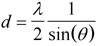
For both form and structure factors there is an inverse relationship between structural size and the spacing or position in angle of the scattered or diffracted radiation. That is, large structures are seen as small angles relative to the incident beam and small structures are seen at high angles. So diffraction from atomic planes or scattering from individual atoms are seen in the diffraction regime with the diffraction angle larger than 6 degrees, while colloidal and nano-scale features are seen at angles far below 6 degrees for x-rays of wavelength close to 0.1 nm. This inverse relationship between structural size and scattering angle is reflected in Bragg's law that relates a structural repeat distance, d, with the angle of scattering or diffraction, 2q, where for small angle sin(q) ~ q in radians.

Spherulites: Polymers with a high degree of crystallinity, such
as high density polyethylene, form spherical crystalline aggregates when melt
crystallized. These spherulites
are usually on the order of 10 to 50 micron in diameter but are known to grow
much larger in certain cases particularly in melt crystallized polyesters. The
base crystalline structures in spherulites (as in all polymer crystals) are lamellar
platelets. Polymers crystallize in platelets because of an energy balance
between the difference
in crystallite surface energy between lateral surfaces, where folding of the
chains does not occur, the broader fold surfaces and the bulk enthalpy of
crystallization. Spherulites
are composed of fibrillar
lamellar bundles which grow from a nucleation site in the melt. In some cases,
especially in polyesters, polymer spherulites form bands which have been related to coordinated
crystallite growth at periodic spacings form the nucleation site. A band is a small
region of coordinated and more perfect crystalline growth. The exact
mechanism for banding in spherulitic
growth is not known but it has been observed in inorganic crystals, minerals
(agate), and in certain eutectic alloys in metals as well as being widely seen
in polymer and some low-molecular weight organic crystallites.
The feature of spherulitic bands
important to this lab is their regular spacing about the crystalline nucleus
and constant spacing through out the sample. When spherulites which display bands on the
order of 50Ám (these are in very large spherulites) are irradiated with
collimated, monochromatic laser light (HeNe l = 0.6328 Ám), Bragg diffraction
occurs due to the difference in polarizability (index of refraction) between the more
perfect crystalline structure in the band and the less perfect structure in the
remainder of the spherulitic
crystalline aggregate. Several orders of diffraction can be observed in some
cases (up to 6 orders). This will be verified by comparing the d-spacing
according to Bragg's law with observation of these spherulitic bands using optical
microscopes. (Note: Usually spherulites
are described as 3-d spheres but in this experiment a thin film is used where
the "spherulites"
are really disks.)
APPROACH:
Each group will be given a thin sample of a
50:50 blend of isotactic
polyhydroxybutryate
(PHB) and atactic
PHB. This system displays band spacings on the order of 50 Ám. By shining a laser beam
through the sample diffraction rings can be observed. (Note: isotactic PHB
crystallizes, atactic
PHB doesn't.) The presence of the non-crystallizing atactic PHB enhances banding. Optical
diffraction from a human hair and a diffraction grating will also be
investigated.
EQUIPMENT AND SUPPLIES:
1) Banded spherulitic PHB
samples between glass cover slips. BE CAREFUL TO HOLD THE SAMPLES ONLY BY THEIR
EDGES SO FINGERPRINTS DO NOT INTERFERE WITH THE EXPERIMENT.
2.) HeNe Laser. DO NOT
SHINE THE LASER BEAM DIRECTLY AT YOUR EYE. BE CAREFUL OF STRAY REFLECTIONS FROM
THE SAMPLE.
3.) A pinhole (100 micron pinhole from Newport Analytical for instance or you may want to try to use a sheet
of white cardboard with a hole punched in the middle for transmission of the
main beam.
4.) Optical
microscope with grid for size measurement.
5.)
Two chemistry mounts to hold the
sample and the cardboard.
6.) Ruler and
pencil.
7.) Human hair
(there should be an ample supply in the lab).
8.) Optical
diffraction grating.
PROCEDURE:
2.) Find a
position where fairly complete rings are observed and trace the rings on the
white cardboard. Record the distance from the sample to the cardboard.
3.) Repeat 2 for
another position in the sample where the diffraction rings are of different
diameter.
4.) Observe the
sample in the optical microscope. Measure several band spacings (at least 10). Also record the maximum
and minimum band spacings.
5.) Observe the spherulites under
crossed polars
and note their appearance.
6.) Repeat
experiment for several human hairs of different color and coarseness and for
the diffraction grating.
RESULTS:
1.) Calculate the
diffraction angle, 2q, using the sample to cardboard distance and the ring
radius.
2.) Use Bragg's
law to calculate the d-spacing for the diffraction ring.
3.) If higher
order rings are observed repeat 3 and 4 with "n" in Bragg's law equal
to the integer order of reflection.
4.) Using the
microscopy data calculate a mean and standard deviation for the band spacing.
5.) Do a similar
analysis for human hair and a diffraction grating.
REPORT:
1.) How do the
band spacings
observed using microscopy compare with the results using Bragg's law?
2.) As you scan
the laser across the sample regions of partial arcs and close to full arcs
(rings) are seen in the diffraction pattern. This is because the laser beam
does not irradiate the entire spherulite
but only a part of the spherulite.
It has been noticed that the larger the spherulites the smaller the arcs which
are observed. Explain this effect.
3.) Other than
partial arcs and whole diffraction rings the diffraction pattern sometimes
takes other shapes. Describe these shapes. What might give rise to these shapes?
4.) In most common
x-ray diffractometers
a line trace through the main beam (across the pattern you observed in 2-d) is
taken. Sketch the appearance of such a linear detector pattern for this system.
5.) If partial
arcs were observed it would be possible to miss the diffraction rings using a linear
detector. How could this be circumvented? How does this relate to the
measurement of an oriented sample in XRD using a line beam profile?
6.) Comment on the
difference in the type of information which can be obtained using diffraction
and microscopy. In a study of
banding at different crystallization temperatures which would yield more useful
information?
7.) What do the
diffraction peaks from human hair correspond to?
8.) How do the diffraction
patterns form human hair and the diffraction grating compare with those from
the polymer spherulitic
bands?